孤立性肠系膜上动脉夹层(isolated superior mesenteric artery dissections,ISMAD)是临床上较少见的血管外科疾病,具有起病急、发展快和误诊率高等特点[1]。ISMAD临床表现主要为急性剧烈腹痛,脐周痛常见,由于肠系膜上动脉(superior mesenteric artery,SMA)真腔受压,肠管缺血,严重时可致腹膜炎[2]。ISMAD的危害在于直接引起肠缺血甚至肠坏死,并存在夹层动脉瘤破裂的风险。该病症状不典型,病因尚未明确,其治疗方案包括保守治疗、腔内治疗和外科手术,目前尚未统一[3-5]。及时进行保守治疗往往能取得较好的效果[6]。本研究通过回顾性分析我院2012年1月—2017年3月收治的37例ISMAD患者的临床资料,其中18例仅行保守治疗后好转,其余19例先行保守治疗失败后继行腔内治疗,对比两组资料从而探讨ISMAD保守成功的预测因素,现总结报告如下。
1 资料与方法
1.1 一般资料
2012年1月—2017年3月共有37例ISMAD患者接受治疗。其中男34例,女3例;年龄38~92岁,平均(54.1±11.4)岁。主要合并症包括高血压11例、糖尿病2例、房颤史3例、冠心病2例。所有患者均以腹痛为主要症状收住入院,体检均无腹膜刺激征,且全部由计算机断层扫描血管造影术(computed tomography angiography,CTA)明确诊断。诊断明确后,所有患者先接受保守治疗,其中18例行保守治疗后腹痛症状缓解或消失,定义该组为保守成功组;其余19例行保守治疗失败,腹痛无法缓解甚至加重且无腹膜刺激征,改行腔内治疗,定义该组为保守失败组。该研究未有因腹膜刺激征急诊行剖腹探查病例。
病例纳入标准:入院经CTA检查诊断ISMAD。病例排除标准:既往腹部手术史、肠系膜上动脉行介人手术史和非ISMAD引起的肠系膜上动脉栓塞。
1.2 治疗方法
1.2.1 保守治疗 禁食水、肠外营养支持、扩血管、抗凝及抗血小板治疗等;合并高血压者控制血压到合理的水平[130/80mmHg(1 mmHg=0.133 kPa)]以下。期间密切观察患者,如果腹痛症状缓解,则继续保守治疗。
1.2.2 腔内治疗 经股动脉(18例)或桡动脉(1例)途径将Cobra导管插至SMA开口,用0.035交换导丝通过夹层段后,将导丝插入回肠动脉分支。固定导丝,撤出导管。 交换长鞘至SMA开口,单弯导管配合泥鳅导丝仔细选入SMA远端,反复造影确定导管位于真腔。交换Supracore导丝,在导丝引导下将自膨式裸支架送至夹层部位,确认支架到位后,缓慢释放支架,必要时用球囊进行后扩张。
所有患者顺利出院后继续口服阿司匹林100 mg/d加氯吡格雷75 mg/d,6个月后行CTA复查。
1.3 预测因素分析
根据患者的一般临床资料及CTA图像,提取保守成功组及保守失败组中患者的相关因素,包括患者年龄、病程、夹层距离(定义:夹层假腔近端离肠系膜上动脉根部的距离)、夹层长度(定义:整个夹层假腔的长度)、腹主动脉-肠系膜上动脉夹角(superior mesenteric artery-distal aorta angle,SAA:在肠系膜上动脉和主动脉分叉水平,分别沿主动脉前壁和肠系膜上动脉下壁作切线,两者之间的夹角)及真腔狭窄度[定义:(夹层近端管腔直径–最狭窄处管腔直径)/夹层近端管腔直径×100%]。通过比较两组患者中的相关因素,筛选出有统计学差异的因素即为影响ISMAD保守治疗效果的预测因素。
1.4 统计学处理
采用SPSS 19.0软件统计分析,计数资料用频数及百分率[n(%)]表示,组间比较采用χ2检验;计量资料用均值±标准差( ±s)表示,组间比较采用t检验,P<0.05为差异有统计学意义。
2 结 果
2.1 两组治疗情况

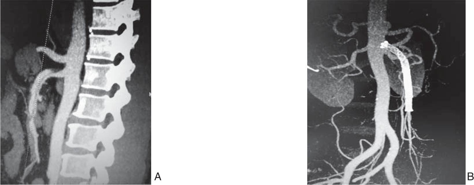
图1 ISMAD患者腔内治疗前后CTA影像学表现 A:术前CTA示SMA真腔细小,部分造影剂通过破口进入假腔;B:腔内治疗6个月后复查CTA示SMA内2枚支架在位,假腔消失,SMA血流通畅
Figure 1 CTA images of ISMAD patients before and after endovascular treatment A: Preoperative CTA showing narrow true lumen of the SMA and some contrast medium entering into the false lumen through the tear; B: Review CTA on 6 months after endovascular treatment showing no displacement of the two stents, disappearance of the false lumen and patent SMA
保守成功组所有患者经保守治疗4~6 d后均顺利出院。保守失败组19例患者腔内治疗均顺利完成,所有患者根据造影情况都植入了自膨式裸支架,规格均在(5~8)×(60~80)mm范围,平均手术时间为(85±42)min。所有19例患者于腔内治疗后3~5 d腹痛好转顺利出院。37例患者出院后随访3~24个月,中位随访15个月,有2例曾经保守治疗好转的患者因腹痛再次入院行保守治疗后顺利出院,其余患者均无明显腹部不适症状;腔内治疗的患者复查CTA示支架在位,假腔消失,肠系膜上动脉显示良好(图1)。
2.2 两组相关因素比较
两组患者的一般临床资料及CTA图像测量数据见表1。两组在年龄、病程方面差异无统计学意义(均P>0.05)。保守成功组SAA≤75°的患者比例明显少于保守失败组(27.8% vs. 63.2%,P<0.05);保守成功组夹层距离明显大于保守失败组[(2.34±0.75)cm vs.(1.47±0.74)cm,P<0.01];保守成功组夹层长度明显小于保守失败组[(3.13±1.17)cm vs.(4.57±2.62)cm,P<0.05];保守成功组真腔狭窄度明显小于保守失败组[(33.9±16.7)% vs.(65.8±22.9)%,P<0.01]。
表1 两组患者相关因素比较
Table 1 Comparison of the relevant factors between the two groups of patients

因素 保守成功组(n=18) 保守失败组(n=19) t/χ2 P年龄(岁, ±s) 53.7±9.0 54.5±13.5 -0.20 >0.05病程≤48 h [n(%)] 12(66.7) 12(63.2) 0.05 >0.05 SAA≤75°[n(%)] 5(27.8) 12(63.2) 4.65 <0.05夹层距离(cm,
 ±s) 2.34±0.75 1.47±0.74 3.52 <0.01夹层长度(cm,
 ±s) 3.13±1.17 4.57±2.62 2.14 <0.05真腔狭窄度(%,
 ±s) 33.9±16.7 65.8±22.9 -4.83 <0.01
3 讨 论
ISMAD临床少见,其发病机制尚不明确。有文献[7]报道,ISMAD好发于中年男性,高血压、糖尿病、血管炎、动脉粥样硬化、动脉囊性中膜坏死,结缔组织病及自身免疫性疾病是高危因素。本组患者中男34例,女3例,平均年龄54.1岁,与文献[8]报道相似。ISMAD的主要临床表现为腹痛,其他可能出现的症状包括腹胀、恶心、呕吐、便血等,这与SMA 真腔狭窄受压导致的肠管肠壁缺血以及夹层周围炎症的反应刺激内脏神经丛有关。
ISMAD的治疗包括保守治疗、腔内治疗和外科手术。在过去,外科手术是治疗本病的常规手段。手术方式较多,如旁路搭桥、内膜切除、夹层动脉瘤切除人造血管移植等[9]。虽然外科手术疗效确切,但是由于其具有手术难度高、风险和创伤大、术后恢复慢等缺点,以及血管腔内修复技术的兴起和保守治疗的认识与发展,外科手术治疗ISMAD的比例总体呈下降趋势。若发生肠缺血坏死,则外科手术切除坏死肠管是唯一选择[10]。对于ISMAD选择何种治疗方法,目前国内外尚无统一意见,且存在较大争议。保守治疗的目的是预防SMA血栓形成闭塞,避免出现肠缺血。只要保守治疗及时充分,多数ISMAD患者可获得治愈[11]。但也有不少患者在保守治疗后症状无明显缓解或加重时,则需要考虑手术治疗或腔内治疗[12]。故如何预测保守治疗的效果,目前尚未有人研究。本研究发现,当SAA>75°、夹层距离较长、夹层长度较小及真腔狭窄率较小时,保守治疗效果偏向良好。另外,年龄大小与病程长短并不影响保守治疗的效果。
保守治疗成功与否往往与患者病情的严重程度相关。由于症状严重程度评价标准不统一,无法横向比较,故通过患者的CTA影像学特点来评估病情的严重程度较为科学。Park等[13]认为肠系膜上动脉弯曲的程度和ISMAD的发生具有一定关系,通过血液动力学的初步研究,发现随着SAA大小的变化,肠系膜上动脉中血液动力学参数也随之变化。部分研究[14-15]发现随着SAA的增大,患ISMAD的风险逐渐增大,SAA和ISMAD的发生具有相关性。本研究发现SAA与保守治疗的效果相关,当SAA>75°时,保守治疗效果较好。
依据夹层破裂口的位置及管腔内是否伴有血栓,Yun等[16]将其分为3型:Ⅰ型,假腔有入口和出口,血流通畅;IIa型,血流可经入口进入假腔,但无出口;IIb型,假腔内血栓形成,无血流;III型,夹层导致真、假腔均闭塞。并且该作者发现ISMAD的症状严重程度与其提出的分型无明显关系。Luan等[17]提出一种基于夹层长度和位置的影像学分型方法:局限于肠系膜上动脉弯曲部为B型,向近端延展为A型,向远端延展为C型(限于肠系膜上动脉主干)、D型(累及回结肠动脉或远端回肠动脉)。笔者[18]发现真腔狭窄越严重、夹层近端距主动脉越近、夹层长度越长,临床症状越严重。本研究发现夹层距离越长、夹层长度越小及真腔狭窄度越小时,保守治疗效果越好。
保守治疗失败后,若无腹膜刺激征,可转为腔内治疗。腔内治疗目的是恢复SMA真腔血流,促进夹层重塑,有利于快速缓解症状和恢复正常的肠功能。由于其具有有效、创伤小、恢复快等特点,腔内治疗已成为ISMAD除保守治疗外的首选治疗方法[19]。本组患者19例行腔内治疗,均顺利植入裸支架,患者腹痛症状好转。复查CTA发现19例患者SMA真腔通畅,所有支架均通畅,无血栓形成,假腔血栓形成并缩小,效果满意。但腔内技术治疗ISMAD也存在一些缺点,例如对血管条件要求高(血管直径要适宜,夹层范围不宜过大)、手术费用高、缺乏长期随访的数据资料等[20]。
综上所述,SAA>75°、夹层距离较长、夹层长度较小及真腔狭窄度较小是ISMAD保守治疗成功的预测因素。当保守治疗失败时,可选择安全有效的腔内治疗。但本组病例数较少,结果尚需更多的临床资料证实。
参考文献
[1] 贾中芝, 田丰, 李绍钦, 等. 孤立性肠系膜上动脉夹层的分型、影像诊断及其预后[J]. 中华胸心血管外科杂志, 2013, 29(3):143–146. doi:10.3760/cma.j.issn.1001–4497.2013.03.006.Jia ZZ, Tian F, Li SQ, et al. The type imaging diagnosis and prognosis of isolated superior mesenteric artery dissection[J].Chinese Journal of Thoracic and Cardiovascular Surgery, 2013,29(3):143–146. doi:10.3760/cma.j.issn.1001–4497.2013.03.006.
[2] Subhas G, Gupta A, Nawalany M, et al. Spontaneous isolated superior mesenteric artery dissection: a case report and literature review with management algorithm[J]. Ann Vasc Surg, 2009,23(6):788–798. doi: 10.1016/j.avsg.2008.12.006.
[3] Jibiki M, Inoue Y, Kudo T. Conservative treatment for isolated superior mesenteric artery dissection[J]. Surg Today, 2013,43(3):260–263. doi: 10.1007/s00595–012–0304–8.
[4] Leung DA, Schneider E, Kubik-Huch R, et al. Acute mesenteric ischemia caused by spontaneous isolated dissection of the superior mesenteric artery: treatment by percutaneous stent placement[J].Eur Radiol, 2000, 10(12):1916–1919.
[5] Katsura M, Mototake H, Takara H, et al. Management of spontaneous isolated dissection of the superior mesenteric artery:Case report and literature review[J]. World J Emerg Surg, 2011,6:16. doi: 10.1186/1749–7922–6–16.
[6] Garrett HE Jr. Options for treatment of spontaneous mesenteric artery dissection[J]. J Vasc Surg, 2014, 59(5):1433–1439. doi:10.1016/j.jvs.2014.01.040.
[7] Sakamoto I, Ogawa Y, Sueyoshi E, et a1. Imaging appearances and management of isolated spontaneous dissection of the superior mesenteric artery[J]. Eur J Radiol, 2007, 64(1):103–110.
[8] Wu XM, Wang TD, Chen MF. Pereutaneous endovascular treatment for isolated spontaneous superior mesenteric artery dissection:report of two cases and literature review[J]. Catheter Cardiovasc Interv, 2009, 73(2):145–151. doi: 10.1002/ccd.21806.
[9] 栾景源, 李选. 孤立性肠系膜上动脉夹层国内现状及展望[J]. 中国微创外科杂志, 2016, 16(10):865–869. doi:10.3969/j.issn.1009–6604.2016.10.001.Nuan JY, Li X. Domestic Situation and Future Perspective of Isolated Dissection of the Superior Mesenteric Artery[J]. Chinese Journal of Minimally Invasive Surgery 2016, 16(10):865–869.doi:10.3969/j.issn.1009–6604.2016.10.001.
[10] 许永乐, 熊江, 郭伟, 等. 孤立性肠系膜上动脉夹层的治疗策略[J]. 介入放射学杂志, 2010, 19(9):694–697. doi:10.3969/j.issn.1008–794X.2010.09.008.Xu YL, Xiong J, Guo W, et al. Therapeutic strategy for the treatment of isolated spontaneous dissection of superior mesenteric arteries[J]. Journal of Interventional Radiology, 2010, 19(9):694–697. doi:10.3969/j.issn.1008–794X.2010.09.008.
[11] Park YJ, Park KB, Kim DI, et a1. Natural history of spontaneous isolated superior mesenteric artery dissection derived from followup after conservative treatment[J]. J Vasc Surg, 2011, 54(6):1727–1733. doi: 10.1016/j.jvs.2011.07.052.
[12] Li N, Lu QS, Zhou J, et a1. Endovascular stent placement for treatment of spontaneous isolated dissection of the superior mesenteric artery[J]. Ann Vasc Surg, 2014, 28(2):445–451. doi:10.1016/j.avsg.2013.01.028.
[13] Park YJ, Park CW, Park KB, et a1. Inference from clinical and fluid dynamic studies about underlying cause of spontaneous isolated superior mesenteric artery dissection[J]. J Vasc Surg, 2011,53(1):80–86. doi: 10.1016/j.jvs.2010.07.055.
[14] 张弘, 吴忠隐, 熊江, 等. 腹主动脉-肠系膜上动脉夹角在自发性孤立性肠系膜上动脉夹层中的意义[J]. 中华普通外科杂志, 2016,31(2):134–136. doi:10.3760/cma.j.issn.1007–631X.2016.02.015.Zhang H, Wu ZY, Xiong J, et al. The significance of the angle between the superior mesenteric artery and aorta in spontaneous isolated superior mesenteric artery dissection[J]. Zhong Hua Pu Tong Wai Ke Za Zhi, 2016, 31(2):134–136. doi:10.3760/cma.j.issn.1007–631X.2016.02.015.
[15] Kim YW. Current Understandings of Spontaneous Isolated Superior Mesenteric Artery Dissection[J]. Vasc Specialist Int, 2016,32(2):37–43. doi: 10.5758/vsi.2016.32.2.37.
[16] Yun WS, Kim YW, Park KB, et a1. Clinical and angiographic follow-up of spontaneous isolated superior mesenteric artery dissection[J]. Eur J Vasc Endovasc Surg, 2009, 37(5):572–577. doi:10.1016/j.ejvs.2008.12.010.
[17] Luan JY, Guan X, Li X, et al. Isolated superior mesenteric artery dissection in China[J]. J Vasc Surg, 2016, 63(2):530–536. doi:10.1016/j.jvs.2015.09.047.
[18] 栾景源, 李选, 许佳文, 等. 增强CT诊断孤立性肠系膜上动脉夹层[J]. 中国介入影像与治疗学, 2014, 11(8):515–519.Nuan JY, Li X, Xu JW, et al. Contrast-enhanced CT in diagnosis of isolated dissection of the superior mesenteric artery[J]. Chinese Journal of Interventional Imaging and Therapy, 2014, 11(8):515–519.
[19] 周建国, 李栋林, 何杨燕, 等. 自发性孤立性肠系膜上动脉夹层的腔内治疗[J]. 中华普通外科杂志, 2015, 30(12):949–952.doi:10.3760/cma.j.issn.1007–631X.2015.12.006.Zhou JG, Li DL, He YY, et al. Endovascular treatment of spontaneous isolated dissection of the superior mesenteric artery[J].Zhong Hua Pu Tong Wai Ke Za Zhi, 2015, 30(12):949–952.doi:10.3760/cma.j.issn.1007–631X.2015.12.006.
[20] 张敏宏, 郭伟. 孤立性肠系膜上动脉夹层的诊疗策略[J]. 中国血管外科杂志:电子版, 2015, 7(4):220–222. doi:10.3969/j.issn.1674–7429.2015.04.004.Zhang MH, Guo W. Strategies for diagnosis and treatment of isolated dissection of the superior mesenteric artery [J]. Chinese Journal of Vascular Surgery: Electronic Version, 2015, 7(4):220–222. doi:10.3969/j.issn.1674–7429.2015.04.004.