乳腺癌是我国女性最常见的恶性肿瘤,是全球女性癌症相关死亡的主要原因,其发病率近年来逐渐升高,且有年轻化趋势[1-2]。一项统计显示,我国2013年新发乳腺癌约278 800例,占新发癌症病例的17.07%,且城市居民的发病率高于农村,发病高峰在55~60岁,乳腺癌在我国癌症死亡原因中位列第五[3]。目前,手术切除是乳腺癌的首选治疗,而腋窝淋巴结(axillary lymph node,ALN)状态则被认为是乳腺癌重要的预后因素[4],因此治疗期间对ALN状态的评估十分关键。过去曾将腋窝淋巴结清扫术(axillary lymph node dissection,ALND)作为乳腺癌患者的标准治疗,但ALND可能会导致肢体疼痛及麻木、淋巴水肿、肩部活动受限等并发症[5],前哨淋巴结活检(sentinel lymph node biopsy,SLNB)的应用,使前哨淋巴结(sentinel lymph node,SLN)阴性患者免行ALND,减少了并发症的发生,提高了生活质量,对于临床淋巴结阴性(clinically node negative,cN0)的早期乳腺癌患者可作为首选治疗方案[6]。SLNB成功与否,和示踪剂的选择有密切联系。亚甲蓝和99mTc硫胶体是目前临床上应用最多的两种示踪剂,两者联合应用可提高SLN检出率,并降低假阴性率[7]。近年来的一些研究中使用吲哚菁绿(indocyanine green,ICG)、超顺磁氧化铁(superparamagnetic iron oxide,SPIO)、靛胭脂、纳米炭等新型示踪剂代替传统的示踪剂,或与传统示踪剂联合应用,获得较理想的检出率和假阴性率[8-10],但这些示踪剂距离广泛临床应用仍有一段距离。国内较多研究对比了ICG联合亚甲蓝与单用亚甲蓝在SLNB中的应用价值,但这些研究纳入的病例数有限,故本研究旨在运用Meta分析的方法对国内相关研究进行汇总分析,以期为ICG联合亚甲蓝示踪法在国内乳腺癌SLNB中的应用提供循证医学依据。
1 资料与方法
1.1 检索策略
在中国知网数据库、万方数据库、维普数据库、PubMed和Cochrane Library中检索得到已公开发表的相关文献。中文检索词有:前哨淋巴结活检、吲哚菁绿、荧光、亚甲蓝、美蓝。英文检索词有:Sentinel Lymph Node Biopsy、Sentinel Node Biopsy、SLNB、Indocyanine Green、Fluorescence、Methylene Blue。检索时间均为自建库至2019年8月。
1.2 文献纳入与排除标准
1.2.1 文献纳入标准 ⑴ 病例均来自中国的医疗机构;⑵ 研究纳入病例均经细胞学或组织病理学确诊为乳腺癌;⑶ 观察组示踪剂为ICG联合亚甲蓝,对照组示踪剂为单用亚甲蓝,或有多个对照组,其中包括单用亚甲蓝组;⑷ 病例均为cN0,无远处转移,无手术禁忌证;⑸ 文中可提取或推算出以下指标中的部分或全部:SLN检出率、检出SLN数、假阴性率、灵敏度、特异度;⑹ 文献发表语言为中文或英文。
1.2.2 文献排除标准 ⑴ 对照组示踪剂非单用亚甲蓝,或未设立对照组;⑵ SLNB术前接受新辅助化疗(neoadjuvant chemotherapy,NAC)或乳腺区放疗;⑶ 无法获取全文或无有效数据的文献;⑷ 文献数据有明显错误或语言指向不明;⑸ 发表语言既非中文也非英文的文献;⑹ 会议论文、学位论文、综述论文。
1.3 文献筛选与数据提取
由2名研究者分别依据纳入和排除标准进行文献资料的筛选,并进行相关数据的提取,对结果进行交叉核对,若遇分歧,则通过协商达成一致或征求第3名研究者的意见后决定是否纳入。提取的数据有:第一作者姓名;文献发表年份;观察组及对照组的样本数、平均年龄;研究病例的临床分期;研究是否涉及自身对照;病例是否均进行ALND;观察组及对照组的相关评价指标。
1.4 文献质量评价
使用Quality Assessment of Diagnostic Accuracy Studies(QUADAS)质量评价工具对纳入文献质量逐一进行评价,共14项条目,文献符合某一条目的标准则记为“是”,未达标准记为“否”,无法评价记为“不清楚”。
1.5 统计学处理
采用Cochrane协作网提供的Review Manager 5.3软件进行统计分析。SLN检出率、假阴性率、灵敏度和特异度的对比结果用比值比(odds ratio,OR)及其95%可信区间(confidence interval,CI)表示,SLN检出数的对比结果用标准化均数差(standard mean difference,SMD)及其95% CI表示。各项研究间的异质性采用I2检验进行分析。若I2≥50%且P≤0.1,则认为研究间异质性较高,采用随机效应模型分析;若I2<50%且P>0.1,则认为研究间异质性较低,采用固定效应模型分析。通过漏斗图评价发表偏倚。P<0.05为差异有统计学意义。
2 结 果
2.1 文献检索和筛选流程
通过中英文数据库检索相关文献,初步检出81篇文献,根据纳入和排除标准逐步筛选,最终纳入15篇文献[11-25],其中中文文献12篇,英文文献3篇,均为国内研究。文献检索和筛选流程见图1。
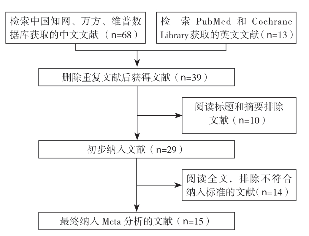
图1 文献检索和筛选流程
Figure1 literature search and screening process
2.2 纳入研究的基本特征
纳入研究的基本特征见表1。表中可见,纳入的15项研究中,2项采用自身对照,绝大多数病例均为I~II期,仅有1项研究为I~III,1项研究纳入病例临床分期不清楚,此外,4项研究无法判断是否所有病例均行ALND。
表1 纳入研究的基本特征
Table1 General features of the included studies
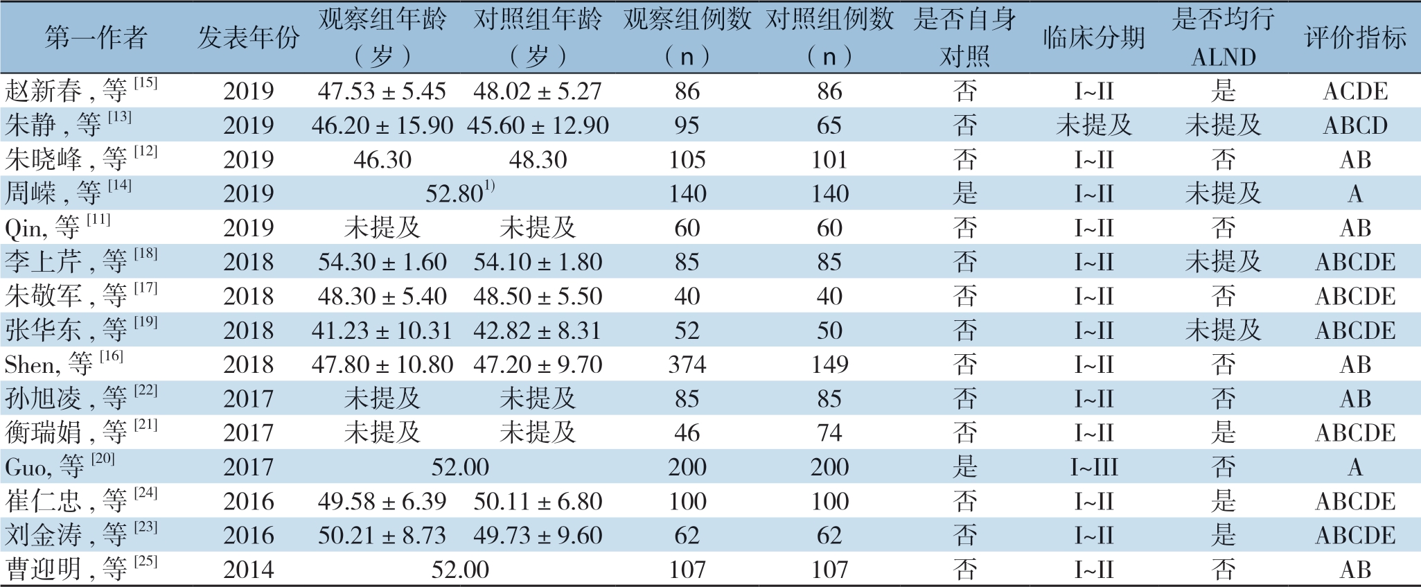
注:1)中位年龄;A:检出率;B:SLN检出数;C:假阴性率;D:灵敏度;E:特异度
Note:1) Median age; A:Detection rate; B:Number of SLNs detected by SLNB; C:False negative rate; D:Sensitivity; E:Specificity
第一作者 发表年份 观察组年龄(岁)对照组年龄(岁)观察组例数(n)对照组例数(n)是否自身对照 临床分期 是否均行ALND 评价指标赵新春,等[15] 2019 47.53±5.45 48.02±5.27 86 86 否 I~II 是 ACDE朱静,等[13] 2019 46.20±15.9045.60±12.90 95 65 否 未提及 未提及 ABCD朱晓峰,等[12] 2019 46.30 48.30 105 101 否 I~II 否 AB周嵘,等[14] 2019 52.801) 140 140 是 I~II 未提及 A Qin,等[11] 2019 未提及 未提及 60 60 否 I~II 否 AB李上芹,等[18] 2018 54.30±1.60 54.10±1.80 85 85 否 I~II 未提及 ABCDE朱敬军,等[17] 2018 48.30±5.40 48.50±5.50 40 40 否 I~II 否 ABCDE张华东,等[19] 2018 41.23±10.31 42.82±8.31 52 50 否 I~II 未提及 ABCDE Shen,等[16] 2018 47.80±10.80 47.20±9.70 374 149 否 I~II 否 AB孙旭凌,等[22] 2017 未提及 未提及 85 85 否 I~II 否 AB衡瑞娟,等[21] 2017 未提及 未提及 46 74 否 I~II 是 ABCDE Guo,等[20] 2017 52.00 200 200 是 I~III 否 A崔仁忠,等[24] 2016 49.58±6.39 50.11±6.80 100 100 否 I~II 是 ABCDE刘金涛,等[23] 2016 50.21±8.73 49.73±9.60 62 62 否 I~II 是 ABCDE曹迎明,等[25] 2014 52.00 107 107 否 I~II 否 AB
2.3 纳入文献质量评价结果
纳入文献质量评价结果见表2。表中条目1~14分别为:疾病谱、选择标准、金标准、疾病进展偏倚、部分参照偏倚、多重参照偏倚、混合偏倚、待评价试验的实施、金标准的实施、试验解读偏倚、金标准解读偏倚、临床解读偏倚、难以解释的试验结果、退出病例。
表2 纳入文献质量评价结果
Table2 Results of quality assessment of the included studies
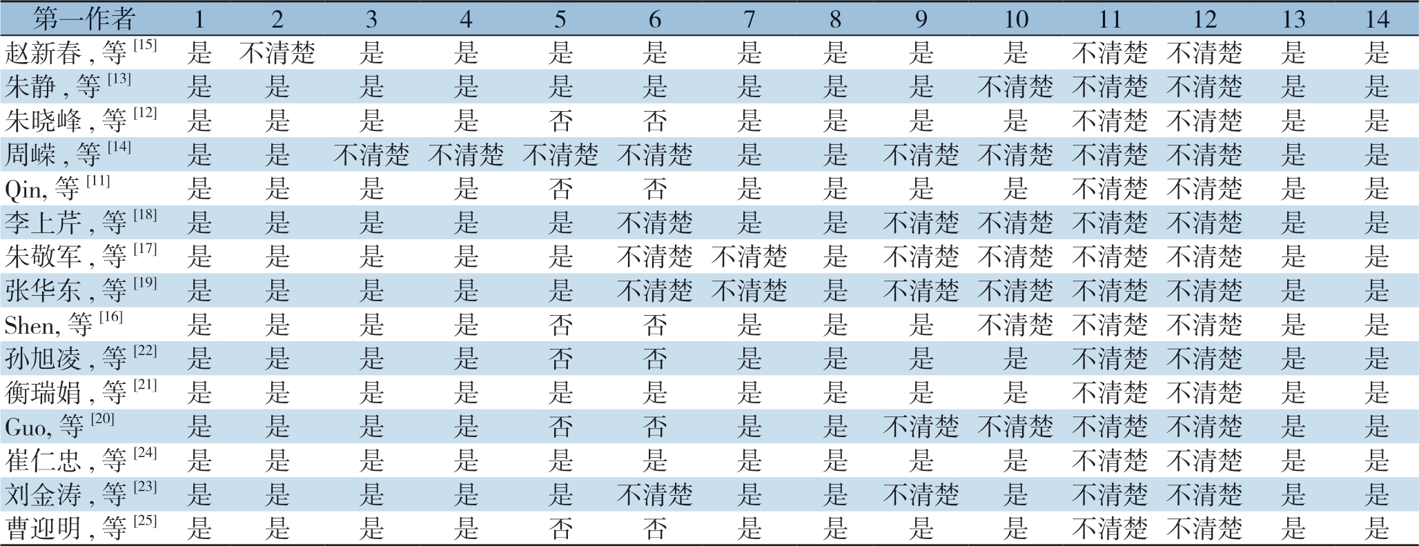
第一作者 1 2 3 4 5 6 7 8 9 10 11 12 13 14赵新春,等[15] 是 不清楚 是 是 是 是 是 是 是 是 不清楚 不清楚 是 是朱静,等[13] 是 是 是 是 是 是 是 是 是 不清楚 不清楚不清楚 是 是朱晓峰,等[12]是 是 是 是 否 否 是 是 是 是 不清楚不清楚 是 是周嵘,等[14] 是 是 不清楚 不清楚 不清楚 不清楚 是 是 不清楚 不清楚 不清楚 不清楚 是 是Qin,等[11] 是 是 是 是 否 否 是 是 是 是 不清楚不清楚 是 是李上芹,等[18] 是 是 是 是 是 不清楚 是 是 不清楚 不清楚 不清楚 不清楚 是 是朱敬军,等[17] 是 是 是 是 是 不清楚 不清楚 是 不清楚 不清楚 不清楚 不清楚 是 是张华东,等[19] 是 是 是 是 是 不清楚 不清楚 是 不清楚 不清楚 不清楚 不清楚 是 是Shen,等[16] 是 是 是 是 否 否 是 是 是 不清楚不清楚不清楚 是 是孙旭凌,等[22]是 是 是 是 否 否 是 是 是 是 不清楚不清楚 是 是衡瑞娟,等[21]是 是 是 是 是 是 是 是 是 是 不清楚不清楚 是 是Guo,等[20] 是 是 是 是 否 否 是 是 不清楚不清楚不清楚不清楚 是 是崔仁忠,等[24]是 是 是 是 是 是 是 是 是 是 不清楚不清楚 是 是刘金涛,等[23] 是 是 是 是 是 不清楚 是 是 不清楚 是 不清楚 不清楚 是 是曹迎明,等[25]是 是 是 是 否 否 是 是 是 是 不清楚不清楚 是 是
2.4 Meta分析结果
2.4.1 SLN检出率 纳入的15项研究[11-25]均报道了SLN检出率,研究间异质性较低(I²=0%,P=0.90),故采用固定效应模型分析。结果提示,ICG联合亚甲蓝组的SLN检出率高于单用亚甲蓝组,差异有统计学意义(OR=5.39,95% CI= 3.46~8.39,P<0.000 01)(图2)。
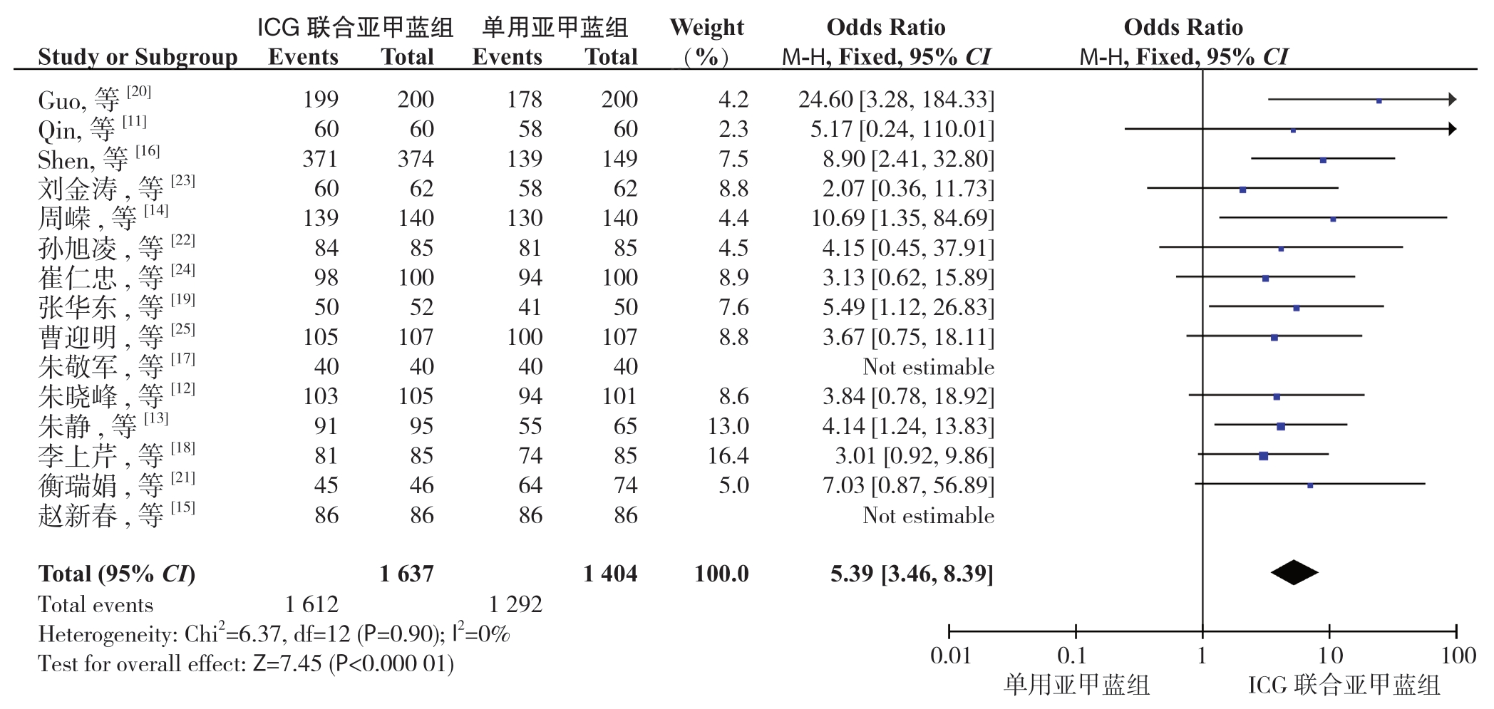
图2 ICG联合亚甲蓝与单用亚甲蓝示踪法B检出率比较
Figure2 Comparison of the detection rates between ICG combined with methylene blue and methylene blue alone
2.4.2 SLN检出数 纳入的15项研究中有 12项[11-13,16-19,21-25]报道了SLN检出数,研究间异质性较高(I²=90%,P<0.000 01),故采用随机效应模型分析。结果提示,ICG联合亚甲蓝组的SLN检出数多于单用亚甲蓝组,差异有统计学意义(SMD=1.06,95% CI=0.76~1.36,P<0.000 01)(图3)。
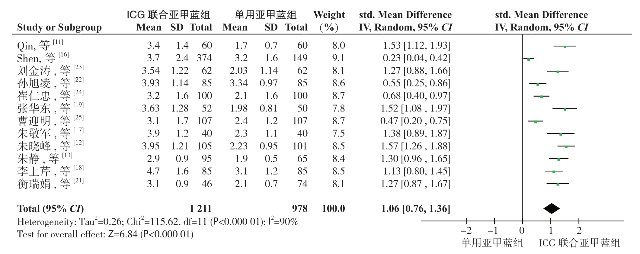
图3 ICG联合亚甲蓝与单用亚甲蓝示踪法SLN检出数(枚)比较
Figure3 Comparison of the number of detected SLNs between ICG combined with methylene blue and methylene blue alone
2.4.3 假阴性率 纳入的15项研究中有 8项[13,15,17-19,21,23-24]报道了或可推算出SLNB假阴性率,研究间异质性较低(I²=2%,P=0.41),故采用固定效应模型分析。结果提示,ICG联合亚甲蓝组的假阴性率低于单用亚甲蓝组,差异有统计学意义(OR=0.15,95% CI=0.08~0.29,P<0.000 01)(图4)。以2018年为界,按文献发表年代将这8项研究分为2个亚组(≥2018年组和<2018年组)进行亚组分析。其中<2018年组的研究间异质性较高(I²=63%,P=0.07),故采用随机效应模型分析以降低偏倚。结果显示,两亚组中ICG联合亚甲蓝组的假阴性率均低于单用亚甲蓝组,差异均有统计学意义(≥2018年组:OR=0.20,95% CI=0.08~0.49,P=0.000 4;<2018年组:OR=0.13,95% CI=0.02~0.93,P=0.04;合 并:OR=0.18,95% CI=0.09~0.36,P<0.000 01)(图5)。
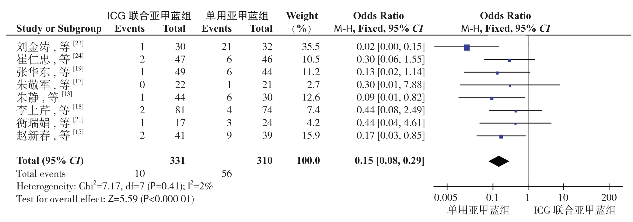
图4 ICG联合亚甲蓝与单用亚甲蓝示踪法用于SLNB假阴性率比较
Figure4 Comparison of the false negative rates of SLNB between ICG combined with methylene blue and methylene blue alone
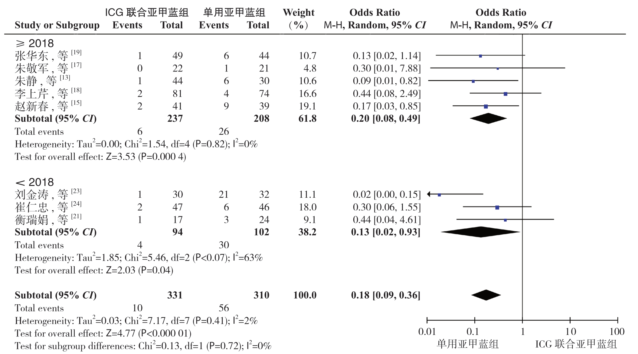
图5 ICG联合亚甲蓝与单用亚甲蓝示踪法用于SLNB假阴性率差异的亚组分析
Figure5 Subgroup analysis of difference in false negative rates of SLNB between ICG combined with methylene blue and methylene blue alone
2.4.4 灵敏度 纳入的15项研究中有8项[13,15,17-19,21,23,24] 报道了或可推算出灵敏度,研究间异质性较低(I²=0%,P=0.86),故采用固定效应模型分析。结果提示,ICG联合亚甲蓝组的灵敏度高于单用亚甲蓝组,差异有统计学意义(OR=5.31,95% CI= 2.68~10.55,P<0.000 01)(图6)。
2.4.5 特异度 纳入的15项研究中有7项[15,17-19,21,23,24]报道了或可推算出特异度,研究间异质性较低(I²=0%,P=0.90),故采用固定效应模型分析。结果提示,ICG联合亚甲蓝组的特异度高于单用亚甲蓝组,差异有统计学意义(OR=2.71,95% CI= 1.35~5.45,P=0.005)(图7)。
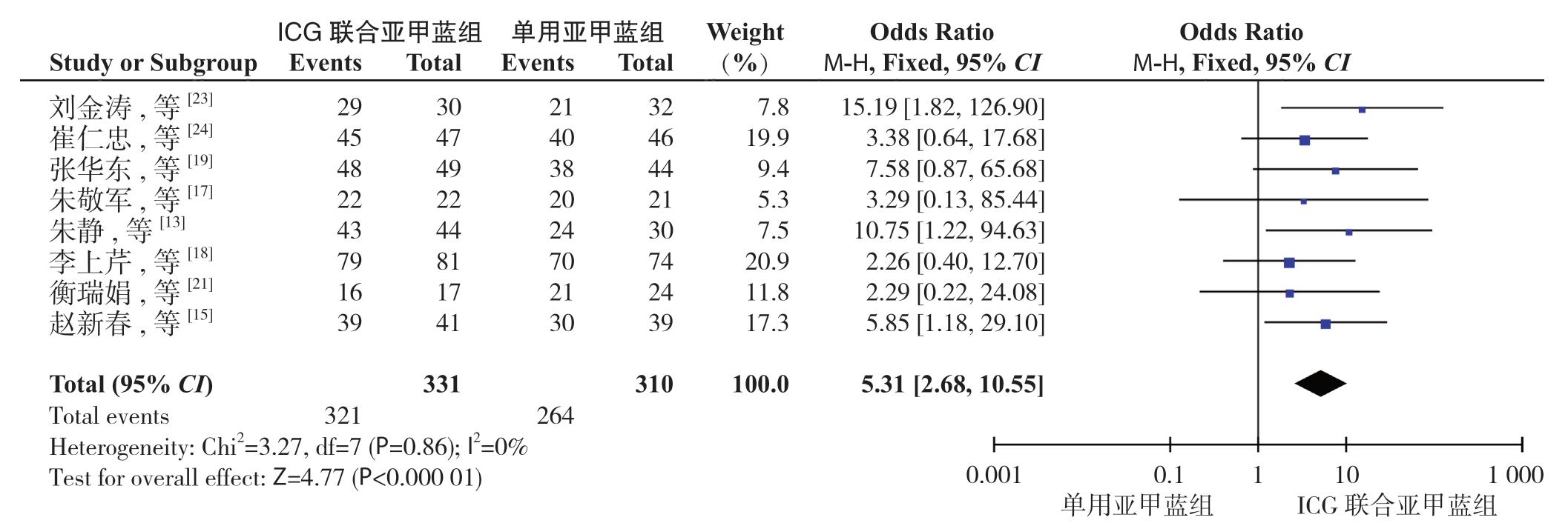
图6 ICG联合亚甲蓝与单用亚甲蓝示踪法用于SLNB灵敏度比较
Figure6 Comparison of the sensitivities between ICG combined with methylene blue and methylene blue alone for SLNB

图7 ICG联合亚甲蓝与单用亚甲蓝示踪法用于SLNB特异度比较
Figure7 Comparison of the specificities between ICG combined with methylene blue and methylene blue alone for SLNB
2.5 发表偏倚评价
本Meta分析中,各项研究指标所纳入的文献篇数不等,故采用报道检出率的15篇文献[11-25]进行发表偏倚的评估。由检出率的漏斗图可以看出,左右两侧基本对称,提示存在发表偏倚可能性较小(图8)。
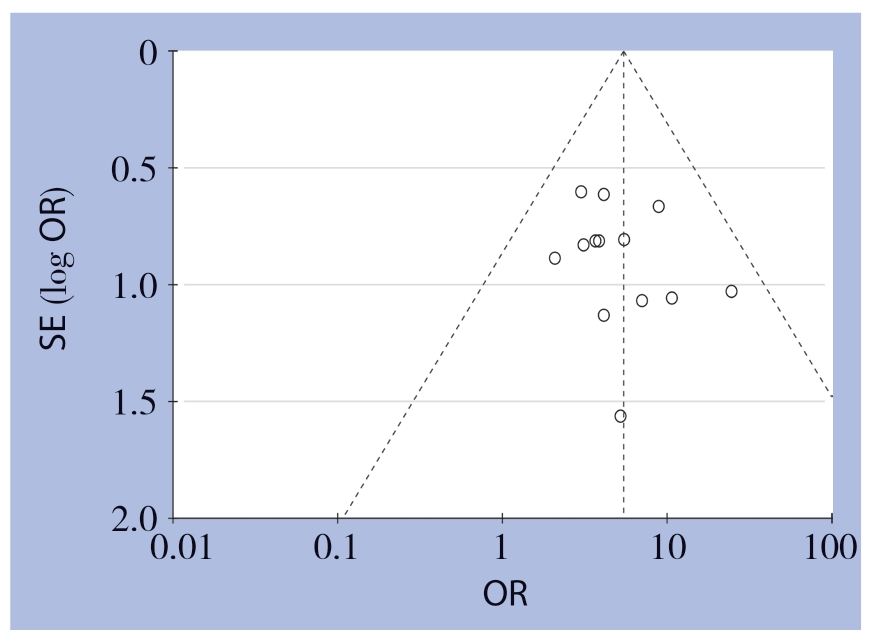
图8 检出率的漏斗图
Figure8 Funnel plot for detection rate
3 讨 论
SLNB已成为多数cN0的早期乳腺癌患者的标准治疗方案,而示踪剂的正确选择对SLNB的成功进行有重要意义。目前临床上使用最多的示踪剂是亚甲蓝和99mTc硫胶体,将两者联合应用,可获得96%的检出率及7.3%的假阴性率[26]。放射性核素可增加辐射暴露的风险,且需强制许可,实用性低,故国内较多医疗机构仍单用亚甲蓝进行SLNB,即使是经验丰富的外科医师,往往成功率和假阴性率也不理想[27]。亚甲蓝和放射性胶体联合应用相比单用其一,虽可提高SLN检出率,降低假阴性率,但尚缺乏充足的临床意义,且增加了腋窝未受累淋巴结的切除率[4]。因此,有学者[8-9,28]对新型示踪剂进行了研究,如ICG、SPIO、靛胭脂、纳米炭等,并证实这些示踪剂具有一定的临床应用价值。
ICG是一种近红外显像剂,波长765 nm的近红外辐射激活ICG分子,发射出波长830 nm的荧光,可被近红外光谱成像系统检测到[29]。Kitai等[30]首次报道了使用吲哚菁绿荧光(indocyanine green fluorescence,ICG-F)和近红外光谱成像系统的SLNB技术,并获得了94%的SLN检出率。后多项研究[29,31]证实ICG用于乳腺癌SLNB的准确性和安全性。ICG目前已被美国食品药品监督管理局(Food and Drug Administration,FDA)和欧洲药品管理局(European Medicines Agency,EMA)批准使用,可有效应用于多种肿瘤的鉴定及特征识别,且可用于淋巴结转移的诊断[32]。
由于单个临床试验的样本量有限,故有学者对ICG在SLNB中的应用价值进行系统评价。Zhang等[33]对ICG用于乳腺癌SLNB中的诊断效能进行Meta分析,共纳入了19项研究,其中6项研究报道了诊断数据,最终得到SLN检出率为98%,合并灵敏度为92%,合并特异度为100%,合并假阴性率为8%,诊断比值比(diagnostic odds ratio,DOR)为311.47,综合受试者工作特性(summary receiver operating characteristic,SROC)曲线下面积(area under the curve,AUC)为0.975 8,这表明ICG应用于SLNB具有良好的诊断效能。另外,Sugie等[34]采用Meta分析的方法将ICG示踪法和放射性核素法进行对比,纳入12项研究,结果得出,两种方法在SLN检出率上无显著差异,但ICG法可能在腋窝分期方面更有优势。后来国内吕莹等[35]又用Meta分析的方法对比了单用ICG与放射性核素在SLNB中的应用效果,共纳入16项研究,最终得出两种方法获得的SLN检出率和假阴性率无显著差异,结果和Sugie等研究[34]的结果部分相似。此外,Goonawardena等[36]的Meta分析在Sugie等研究[34]的基础上进行补充,共纳入19项研究,最终仍得出两法在SLN检出率上无显著差异。这些Meta分析结果说明,ICG有望成为传统示踪剂的替代品。
此外,有学者将ICG与传统示踪剂联用进行临床试验,在SLNB质量上取得一定的改善。例如,Guo等[37]的一项研究纳入了86例乳腺癌患者(Tis 12例,T1 28例,T2 46例),在单用ICG作为示踪剂的基础上,联用亚甲蓝,结果SLN检出率与单用ICG无显著差异,但假阴性率从12%降至4%。另外,Jung等[38]的一项II期随机对照试验纳入86例cN0乳腺癌患者,实验组联合应用ICG、放射性核素和亚甲蓝进行SLNB,对照组单用放射性核素,实验组在SLN平均检出数上显著高于对照组,但在SLN检出率、首个SLN检出时间、SLN平均检出时间和SLNB总时间上无显著差异,但这种多模式联合法(multimodal method,MMM)更适用于NAC后需要进行SLNB的患者。近年来Takemoto等[39]的一项临床研究将ICG和靛胭脂联合,与单用ICG对比,共纳入202例0-IIA期接受保乳治疗的乳腺癌患者,结果联合组的SLN检出率显著高于单用ICG组,尤其对于年龄≥60岁和体质量指数(body mass index,BMI)≥25 kg/m2的患者。
然而,目前对联用多种示踪剂临床价值的汇总研究较少。国内多项研究对比了ICG联合亚甲蓝与单用亚甲蓝作为示踪剂在SLNB中的应用价值,但这些研究所纳入的病例数有限,且尚未进行汇总分析。本研究对国内相关研究进行Meta分析,结果得出,ICG联合亚甲蓝示踪法与单用亚甲蓝相比,在乳腺癌SLNB中能获得更高的检出率、SLN检出数、灵敏度和特异度,以及更低的假阴性率,其中SLN检出数这一指标纳入的研究以及2018年前涉及假阴性率的文献异质性较高,该结论还需进一步考证。这种联合示踪法不仅适用于国内放射性核素示踪剂供应有限的医疗机构,且对外科医生手术经验的要求相对较低,可降低放射性暴露的风险,在国内有潜力成为传统示踪法的替代之一。
本研究存在一定的局限性:⑴ 涉及ICG联合亚甲蓝示踪法的研究主要集中于国内医疗机构,国外几乎无法检索到对比ICG联合亚甲蓝与单用亚甲蓝作为示踪剂在SLNB中应用价值的文献,且仅纳入中文和英文文献,可能忽略了其他非中英文文献,这些因素均可能导致纳入研究不够全面。⑵ 纳入的研究中有部分未对所有病例均行ALND,因此仅能得到SLN检出率和检出数等指标,可获得假阴性率的研究有限。⑶ Meta分析本身是对过去已完成的临床研究进行汇总分析,在某些指标上可能存在一定的临床异质性,导致结果可信度降低。因此,ICG联合亚甲蓝示踪法用于SLNB仍需要大量临床研究数据的支撑,随着研究的深入,未来将纳入更多临床研究进行汇总分析,使该法在将来的临床应用中凸显其优势。
[1] Bray F,Ferlay J,Soerjomataram I,et al.Global cancer statistics 2018:GLOBOCAN estimates of incidence and mortality worldwide for 36 cancers in 185 countries[J].CA Cancer J Clin,2018,68(6):394-424.doi:10.3322/caac.21492.
[2] Chen VC,Lin KY,Tsai YH,et al.Connectome analysis of brain functional network alterations in breast cancer survivors with and without chemotherapy[J].PLoS One,2020,15(5):e232548.doi:10.1371/journal.pone.0232548.
[3] Zuo TT,Zheng RS,Zeng HM,et al.Female breast cancer incidence and mortality in China,2013[J].Thorac Cancer,2017,8(3):214-218.doi:10.1111/1759-7714.12426.
[4] Papathemelis T,Jablonski E,Scharl A,et al.Sentinel lymph node biopsy in breast cancer patients by means of indocyanine green using the Karl Storz VITOM(R) fluorescence camera[J].Biomed Res Int,2018,2018:6251468.doi:10.1155/2018/6251468.
[5] Arisio R,Borella F,Porpiglia M,et al.Axillary dissection vs.no axillary dissection in breast cancer patients with positive sentinel lymph node:a single institution experience[J].In Vivo,2019,33(6):1941-1947.doi:10.21873/invivo.11689.
[6] Mansel RE,Fallowfield L,Kissin M,et al.Randomized multicenter trial of sentinel node biopsy versus standard axillary treatment in operable breast cancer:the ALMANAC Trial[J].J Natl Cancer Inst,2006,98(9):599-609.doi:10.1093/jnci/djj158.
[7] Goyal A,Newcombe RG,Chhabra A,et al.Factors affecting failed localisation and false-negative rates of sentinel node biopsy in breast cancer--results of the ALMANAC validation phase[J].Breast Cancer Res Treat,2006,99(2):203-208.doi:10.1007/s10549-006-9192-1.
[8] Ersoy YE,Kadioglu H.Review of novel sentinel lymph node biopsy techniques in breast cancer patients treated with neoadjuvant chemotherapy[J].Clin Breast Cancer,2018,18(4):e555-559.doi:10.1016/j.clbc.2018.01.004.
[9] Man V,Wong TT,Co M,et al.Sentinel lymph node biopsy in early breast cancer:magnetic tracer as the only localizing agent[J].World J Surg,2019,43(8):1991-1996.doi:10.1007/s00268-019-04977-1.
[10] 邹伟伟,白玉,王希龙,等.吲哚菁绿与纳米碳在乳腺癌前哨淋巴结活检中的对比研究[J].中国普通外科杂志,2016,25(11):1627-1632.doi:10.3978/j.issn.1005-6947.2016.11.018.
Zou WW,Bai Y,Wang XL,et al.Comparison of indocyanine green and carbon nanoparticles for detection of sentinel lymph nodes in breast cancer[J].Chinese Journal of General Surgery,2016,25(11):1627-1632.doi:10.3978/j.issn.1005-6947.2016.11.018.
[11] Qin X,Yang M,Zheng X.Comparative study of indocyanine green combined with blue dye with methylene blue only and carbon nanoparticles only for sentinel lymph node biopsy in breast cancer[J].Ann Surg Treat Res,2019,97(1):1-6.doi:10.4174/astr.2019.97.1.1.
[12] 朱晓峰,曾丽莉,黄亮亮,等.吲哚菁绿联合亚甲蓝示踪法在乳腺癌前哨淋巴结活检术中的应用[J].遵义医学院学报,2019,42(1):80-83.doi:10.3969/j.issn.1000-2715.2019.01.016.
Zhu XF,Zeng LL,Huang LL,et al.Indocyanine green in addition to methylene blue for sentinel lymph node biopsy in patients with early breast cancer [J].Journal of Zunyi Medical University,2019,42(1):80-83.doi:10.3969/j.issn.1000-2715.2019.01.016.
[13] 朱静.吲哚菁绿联合亚甲蓝在乳腺癌前哨淋巴结活检中的应用[J].医学临床研究,2019,36(1):113-115.doi:10.3969/j.issn.1671-7171.2019.01.037.
Zhu J.Application of indocyanine green combined with methylene blue in sentinel lymph node biopsy for breast cancer[J].Journal of Clinical Research,2019,36(1):113-115.doi:10.3969/j.issn.1671-7171.2019.01.037.
[14] 周嵘,黄玥,黄颖,等.吲哚菁绿联合亚甲蓝在乳腺癌前哨淋巴结活检中的应用[J].华夏医学,2019,32(3):96-100.doi:10.19296/j.cnki.1008-2409.2019-03-024.
Zhou R,Huang Y,Huang Y,et al.Application of Indocyanine Green combined with Methylene Blue in sentinel lymph node biopsy of breast cancer[J].Acta Medicinae Sinica,2019,32(3):96-100.doi:10.19296/j.cnki.1008-2409.2019-03-024.
[15] 赵新春.吲哚菁绿与亚甲蓝应用于乳腺癌前哨淋巴结活检中的价值研究[J].医药前沿,2019,9(1):101-102.doi:10.3969/j.issn.2095-1752.2019.01.077.
Zhao XC.Value of using indocyanine green and methylene blue in sentinel lymph node biopsy for breast cancer[J].Health World,2019,9(1):101-102.doi:10.3969/j.issn.2095-1752.2019.01.077.
[16] Shen S,Xu Q,Zhou Y,et al.Comparison of sentinel lymph node biopsy guided by blue dye with or without indocyanine green in early breast cancer[J].J Surg Oncol,2018,117(8):1841-1847.doi:10.1002/jso.25058.
[17] 朱敬军,杨金喆,王哲,等.吲哚菁绿导航技术联合亚甲蓝在乳腺癌前哨淋巴结活检中的临床应用价值研究[J].癌症进展,2018,16(15):1857-1860.doi:10.11877/j.issn.1672-1535.2018.16.15.11.
Zhu JJ,Yang JZ,Wang Z,et al.Clinical value of indocyanine green navigation combined with methylene blue in sentinel lymph node biopsy of breast cancer[J].Oncology Progress,2018,16(15):1857-1860.doi:10.11877/j.issn.1672-1535.2018.16.15.11.
[18] 李上芹,苏国森,程少萍,等.吲哚菁绿联合亚甲蓝在乳腺癌腋窝前哨淋巴结活检中的临床应用价值[J].山西医药杂志,2018,47(19):2306-2308.doi:10.3969/j.issn.0253-9926.2018.19.017.
Li SQ,Su GS,Cheng SP,et al.Clinical value of using indocyanine green combined with methylene blue in axillary sentinel lymph node biopsy of breast cancer[J].Shanxi Medical Journal,2018,47(19):2306-2308.doi:10.3969/j.issn.0253-9926.2018.19.017.
[19] 张华东,刘桂琴,张显峰.荧光显影法联合亚甲蓝染色在早期乳腺癌前哨淋巴结活检中的应用价值探讨[J].安徽医学,2018,39(12):1520-1522.doi:10.3969/j.issn.1000-0399.2018.12.029.
Zhang HD,Liu GQ,Zhang XF.Application value of fluorescence display combined with methylene blue in sentinel lymph node biopsy of early breast cancer[J].Anhui Medical Journal,2018,39(12):1520-1522.doi:10.3969/j.issn.1000-0399.2018.12.029.
[20] Guo J,Yang H,Wang S,et al.Comparison of sentinel lymph node biopsy guided by indocyanine green,blue dye,and their combination in breast cancer patients:a prospective cohort study[J].World J Surg Oncol,2017,15(1):196.doi:10.1186/s12957-017-1264-7.
[21] 衡瑞娟,齐攀,冯跃庆,等.吲哚菁绿联合亚甲蓝与单用亚甲蓝在乳腺癌前哨淋巴结活检中的对比研究[J].河南医学研究,2017,26(17):3073-3075.doi:10.3969/j.issn.1004-437X.2017.17.001.
Heng RJ,Qi P,Feng YQ,et al.Comparison study of indocyanine green joint methylene blue with methylene blue alone for sentinel lymph node biopsy in patients with breast cancer[J].Henan Medical Research,2017,26(17):3073-3075.doi:10.3969/j.issn.1004-437X.2017.17.001.
[22] 孙旭凌,黄桂林,申婧,等.吲哚菁绿联合亚甲蓝在早期乳腺癌前哨淋巴结活检术中的应用价值[J].山东医药,2017,57(9):48-50.doi:10.3969/j.issn.1002-266X.2017.09.015.
Sun XL,Huang GL,Shen J,et al.Application value of indocyanine green combined with methylene blue in sentinel lymph node biopsy of early breast cancer[J].Shandong Medical Journal,2017,57(9):48-50.doi:10.3969/j.issn.1002-266X.2017.09.015.
[23] 刘金涛,郭文斌,孙嘉忆.吲哚菁绿联合美蓝作为淋巴示踪剂在乳腺癌SLNB检测中的价值[J].中国普通外科杂志,2016,25(11):1658-1661.doi:10.3978/j.issn.1005-6947.2016.11.023.
Liu JT,Guo WB,Sun JY.Value of indocyanine green combined with methylene blue as lymph node tracer for SLNB detection in breast cancer[J].Chinese Journal of General Surgery,2016,25(11):1658-1661.doi:10.3978/j.issn.1005-6947.2016.11.023.
[24] 崔仁忠,杨接辉,潘承欣.吲哚菁绿联合亚甲蓝在乳腺癌前哨淋巴结活检术中的应用研究[J].海南医学院学报,2016,22(14):1584-1586.doi:10.13210/j.cnki.jhmu.20160414.012.
Cui RZ,Yang JH,Pan CX.Application of Indocyanine green joint methylene blue for sentinel lymph node biophy in patients with breast cancer[J].Journal of Hainan Medical College,2016,22(14):1584-1586.doi:10.13210/j.cnki.jhmu.20160414.012.
[25] 曹迎明,王殊,郭嘉嘉,等.吲哚菁绿联合美蓝在乳腺癌前哨淋巴结活检术中的应用[J].中华普通外科杂志,2014,29(2):119-122.doi:10.3760/cma.j.issn.1007-631X.2014.02.013.
Cao YM,Wang S,Guo JJ,et al.Combination of ICG and methylene blue for mapping sentinel lymph nodes in early breast cancer patients[J].Zhong Hua Pu Tong Wai Ke Za Zhi,2014,29(2):119-122.doi:10.3760/cma.j.issn.1007-631X.2014.02.013.
[26] Kim T,Giuliano AE,Lyman GH.Lymphatic mapping and sentinel lymph node biopsy in early-stage breast carcinoma:a Metaanalysis[J].Cancer,2006,106(1):4-16.doi:10.1002/cncr.21568.
[27] Tian C,Sun X,Cong B,et al.Murine model study of a new receptor-targeted Tracer for sentinel lymph node in breast cancer[J].J Breast Cancer,2019,22(2):274-284.doi:10.4048/jbc.2019.22.e28.
[28] 郭文斌,高伟,刘金涛,等.纳米碳对乳腺癌腋窝前哨淋巴结活检的应用价值[J].中国普通外科杂志,2012,21(11):1346-1349.
Guo WB,Gao W,Liu JT,et al.Application value of carbon nanoparticle in axillary sentinel lymph node biopsy for breast cancer[J].Chinese Journal of General Surgery,2012,21(11):1346-1349.
[29] Lin J,Lin LS,Chen DR,et al.Indocyanine green fluorescence method for sentinel lymph node biopsy in breast cancer[J].Asian J Surg,2020,pii:S1015-9584(20)30045-2.doi:10.1016/j.asjsur.2020.02.003.[Epub ahead of print]
[30] Kitai T,Inomoto T,Miwa M,et al.Fluorescence navigation with indocyanine green for detecting sentinel lymph nodes in breast cancer[J].Breast Cancer,2005,12(3):211-215.doi:10.2325/jbcs.12.211.
[31] 郭文斌,高伟,刘金涛,等.吲哚菁绿荧光导航技术在乳腺癌前哨淋巴结活检中的应用[J].中国普通外科杂志,2015,24(5):658-662.doi:10.3978/j.issn.1005-6947.2015.05.008
Guo WB,Gao W,Liu JT,et al.Application of indocyanine green fluorescence navigation in sentinel lymph node biopsy for breast cancer[J].Chinese Journal of General Surgery,2015,24(5):658-662.doi:10.3978/j.issn.1005-6947.2015.05.008
[32] Egloff-Juras C,Bezdetnaya L,Dolivet G,et al.NIR fluorescenceguided tumor surgery:new strategies for the use of indocyanine green[J].Int J Nanomedicine,2019,14:7823-7838.doi:10.2147/IJN.S207486.
[33] Zhang X,Li Y,Zhou Y,et al.Diagnostic performance of indocyanine green-guided sentinel lymph node biopsy in breast cancer:a Meta-analysis[J].PLoS One,2016,11(6):e155597.doi:10.1371/journal.pone.0155597.
[34] Sugie T,Ikeda T,Kawaguchi A,et al.Sentinel lymph node biopsy using indocyanine green fluorescence in early-stage breast cancer:a meta-analysis[J].Int J Clin Oncol,2017,22(1):11-17.doi:10.1007/s10147-016-1064-z.
[35] 吕莹,程刚.吲哚菁绿荧光示踪法在进行前哨淋巴结活检术中的应用价值[J].当代医药论丛,2019,17(13):164-166.
Lu Y,Cheng G.Application value of indocyanine green fluorescence tracer in sentinel lymph node biopsy[J].Contemporary Medicine Forum,2019,17(13):164-166.
[36] Goonawardena J,Yong C,Law M.Use of indocyanine green fluorescence compared to radioisotope for sentinel lymph node biopsy in early-stage breast cancer:systematic review and metaanalysis[J].Am J Surg,2020,pii:S0002-9610(20)30069-6.doi:10.1016/j.amjsurg.2020.02.001.[Epub ahead of print]
[37] Guo W,Zhang L,Ji J,et al.Evaluation of the benefit of using blue dye in addition to indocyanine green fluorescence for sentinel lymph node biopsy in patients with breast cancer[J].World J Surg Oncol,2014,12:290.doi:10.1186/1477-7819-12-290.
[38] Jung SY,Kim SK,Kim SW,et al.Comparison of sentinel lymph node biopsy guided by the multimodal method of indocyanine green fluorescence,radioisotope,and blue dye versus the radioisotope method in breast cancer:a randomized controlled trial[J].Ann Surg Oncol,2014,21(4):1254-1259.doi:10.1245/s10434-013-3437-0.
[39] Takemoto N,Koyanagi A,Yasuda M,et al.Comparison of the indocyanine green dye method versus the combined method of indigo carmine blue dye with indocyanine green fluorescence imaging for sentinel lymph node biopsy in breast conservative therapy for stage ≤IIA breast cancer[J].BMC Womens Health,2018,18(1):151.doi:10.1186/s12905-018-0646-5.
