下腔静脉滤器作为预防致命性肺栓塞有效的手段,在临床上已经广泛使用[1-2]。但是滤器永久植入后远期并发症很严重[3-6],所以指南指出应以可取出滤器为首选[7]。可取出滤器的种类繁多,从形态上主要分为两类:锥形滤器和纺锤形滤器。纺锤形滤器因为与下腔静脉壁为面接触,导致内膜增生快,所以纺锤形滤器的回收窗只有2周时间。锥形滤器与下腔静脉只有主腿远端点状固定,内膜包裹速度慢,回收时间窗远远长于纺锤形滤器,所以在临床上占绝对比例。但是锥形滤器附腿均为游离状态,使滤器植入后形态稳定性较差,相比纺锤形滤器容易发生倾斜。另外,由于锥形滤器对介入取出的技术要求比较高,回收时发生医源性倾斜也屡见不鲜。严重倾斜时回收钩贴附在下腔静脉壁上,随植入时间增加,回收钩被内膜包裹于下腔静脉壁内,甚至在应力作用下穿透下腔静脉壁。针对这些回收钩包埋在下腔静脉壁内或穿透下腔静脉壁并且介入失败的患者,传统上只能转化为永久植入的滤器或进行开腹手术,切开下腔静脉回收滤器[8]。滤器永久植入后并发症较多,例如下腔静脉缩窄、闭塞、滤器解体、刺透静脉壁导致严重脏器损伤。开腹手术对患者来说创伤大、并发症多、恢复慢,对患者的身体状况要求高,国际上有个别病例的报道采用腹腔镜进行手术。我中心作为全国血栓防治基地,收治了许多其他医院转来进行治疗的这类患者,自2016年开始尝试采用全腹腔镜下或腹腔镜辅助手术取出滤器,达到良好的治疗效果[9]。
1 资料与方法
1.1 一般资料
回顾性分析我院2016年12月—2019年11月收治15例下腔静脉锥形滤器植入患者的临床资料,其中男12例,女3例;平均年龄(47.7±13.3)岁。置入Celect滤器12例,Denali滤器2例,Option滤器1例。所有置入滤器尝试介入取出均失败,术前D-二聚体为(0.44±0.50)mg/L,所有患者术前均行腹部CT检查进行评估。患者在全身麻醉下行全腹腔镜辅助下滤器取出术。记录手术方式、术中及术后并发症及住院时间等。
1.2 术前CT 评估
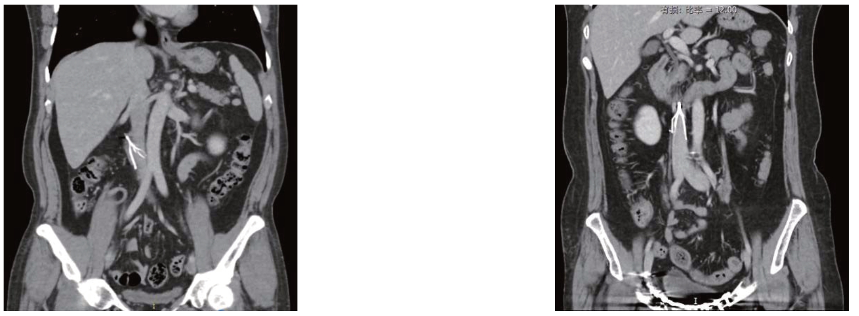
所有患者术前行腹部CT平扫或CT血管造影。所有滤器回收钩均穿透下腔静脉壁,其中1 例滤器回收钩位于左肾静脉上,14例位于右肾静脉以下,1例靠近腹主动脉,1例穿透十二指肠肠壁。CT冠状位显示:回收钩向下腔静脉壁前侧穿出1 例,后侧穿出2 例,右侧穿出7 例,左侧穿出1例,右后侧穿出4例。CT提示回收钩位于下腔静脉壁后侧或右后侧采用腹腔后途径全腹腔镜下滤器取出,回收钩位于下腔静脉壁前方、右侧及左侧采用经腹腔途径(图1-2)。

图1 CT 冠状位显示滤器回收钩穿出下腔静脉壁方向 A:前;B:后;C:左;D:右
Figure 1 Coronal CT images showing the directions of the retrieval hook of the filter penetrating the wall of the inferior vena cava
A:Anterior direction;B:Posterior direction;C:Left direction;D:Right direction

图2 CT 矢状位分别提示Celect 和Denali 滤器回收钩穿透血管壁
Figure 2 Sagittal CT images showing the retrieval hooks of Celect and Denali filters penetrating the wall of the inferior vena cava,respectively
1.3 手术方法
1.3.1 经腹腔途径 在全身麻醉下将患者左侧卧位,于脐、脐与剑突的中点、脐与耻骨联合的中点及脐右侧5 cm、剑突下,分别置人10、10、10、5、5 mm 的Trocar。术中应用腹腔镜器械切开右结肠旁沟外侧腹膜,将升结肠和十二指肠推向内侧,并切开肾周筋膜,显露肾脏、肾静脉及下腔静脉,仔细游离下腔静脉,寻找穿透下腔静脉壁的回收钩。寻找到回收钩后,于回收钩上下约3 mm 处用5-0 Prolene 线分别缝合1 针,应用电钩切开回收钩处静脉壁,完整暴露回收钩,通过Trocar 置入滤器回收鞘,在内镜的直视下应用网篮圈套器圈套回收钩,将滤器回收(图3)。将预先留置的缝合线收紧并打结。彻底止血,留置引流管1 根,取出Trocar,逐层缝合切口。术后给予低分子肝素抗凝(0.4 mL/12 h)治疗。术后引流量<50 mL 拔除引流管。

图3 术中照片 A:腹腔镜直视下滤器回收钩穿出下腔静脉壁;B:应用网篮圈套器回收滤器
Figure 3 Intraoperative views A:Direct sight of the retrieval hook of the filter penetrating the wall of the inferior vena cava under laparoscopy;B:Filter retrieval by using the loop snare technique
1.3.2 经腹膜后途径 全麻下左侧卧位,抬高腰桥,于右肋缘下腋后线、腋前线、腋中线髂嵴上2 cm、髂前上棘上3 cm分别置人10、10、10、5 mm 的Trocar。术中应用腹腔镜器械分离肾蒂,仔细寻找肾动静脉,根据术前CT 所提示回收钩的位置,沿下腔静脉上下分离,可见滤器回收钩,应用电钩仔细游离并完整暴露回收钩,通过Trocar 置入滤器回收鞘,在内镜的直视下应用网篮圈套器圈套回收钩,将滤器回收。应用5-0 Prolene 线缝合下腔静脉破口,防止出血。
1.4 数据处理
计数资料以比例(百分比)[n(%)]表示,计量资料以均数±标准差(x±s)表示。
2 结 果
2.1 患者手术情况
手术方式经腹腔途径9例(60.0%),经腹膜外途径6例(40.0%)。全腹腔镜辅助下滤器成功取出14例(93.3%),其中1例为Option滤器,2例为Denali滤器,11例为Celect滤器。1例(6.7%)Celect滤器腹腔镜手术未能分离回收钩,中转经右侧腹直肌开腹手术成功取出。腹腔镜取出率为93.3%。
2.2 患者围手术期情况
滤器置入时间为平均为(103.9±70.3)d;术中失血给予输血治疗1例(6.7%),切口皮肤感染1例(6.7%);术后平均住院(7.4±2.8)d。所有患者生命体征平稳出院。
3 讨 论
下腔静脉滤器虽然能有效的预防致命性肺栓塞,但是如果植入时间太长或永久植入仍会导致其他并发症发生[10-11]。美国FDA2005年总共收到955例滤器植入后不良事件上报,2010年正式提出在肺栓塞风险降低后尽快取出滤器[12-13]。欧洲CIRSE指南也提出尽量放置可取出滤器[14]。国内指南[15-17]也建议应用可取出滤器。目前,市面上的滤器形态各异,主要可以分为以下几种:(1)纺锤形滤器(以Cordis公司的Optease和国产先健公司为代表);(2)锥形滤器(以COOK公司的Celect和巴德公司的Denali为代表);(3)可转换滤器(以贝朗公司的Convertble为代表);(4)永久滤器。其中前两类是本研究所讨论的可取出滤器,基本是在原永久滤器的基础上增加回收钩从而达到可以取出的目的。纺锤形滤器在下腔静脉内形态稳定,不易发生倾斜。由于纺锤形滤器径向支撑力大,加之与下腔静脉壁呈张力性面接触,内膜增生快,一般在2周左右必须取出,否则滤器和下腔静脉壁粘连严重导致取出失败[18-19]。但是血栓的发展过程最少也需要2~3周才能机化稳定,致命性肺栓塞风险降低,所以纺锤形滤器在临床使用中受到很大制约。锥形滤器在结构上是以支爪的尾端倒钩固定在下腔静脉壁上,形成点接触,以期减少内膜增生问题,延长回收窗。锥形滤器在国外的研究中平均植入时间达180~200 d[20-21],可以进行充分的抗凝、溶栓或消栓的治疗。但是锥形滤器回收窗的延长是以丧失稳定性为代价的,容易发生倾斜[22-24],国内研究[25]报道滤器植入后倾斜率28.2%。回收钩一旦贴壁,会被内膜或血栓包裹,而渐渐深埋于下腔静脉壁内,本组15例患者均为此种情况。同时,不规范的介入取出操作加速了滤器的倾斜和回收钩的穿透。回收钩的穿透有可能导致大出血、腹主动脉穿孔、肠穿孔、下腔静脉缩窄闭塞等一系列并发症[3-4,26]。此时滤器的取出势在必行,原来只能通过开腹手术进行下腔静脉切开[27-28],创伤较大,腹腔镜下滤器取出仅有少量文献报道[29-31]。
CT早期应用在滤器植入后下腔静脉穿透率研究[32],但是血管壁有一定的延展性,CT显示的一、二级穿透仍有可能通过腔内技术取出滤器而不损伤下腔静脉壁。三级以上的穿透,尤其是回收钩的三级穿透则预示着通过介入方法几乎不可能顺利取出滤器。腹腔镜作为微创术式有切口小、损伤小、恢复快的优势,但也存在视野小、对操作技术要求高的弊端。所以术前CT评估手术方案就尤为重要。首先,通过CT可以判断下腔静脉有无血栓形成,只有在下腔静脉通畅的前提下才能进行滤器的取出。其次,通过CT可以明确回收钩穿透下腔静脉壁的位置,如果回收钩偏向前壁穿出,则术式选择经腹腔路径,回收钩偏向后壁穿出则选择腹膜后路径,术式的选择直接影响到手术的难易程度。另外,通过CT可以了解回收钩穿透后下腔静脉周围血肿情况,回收钩穿透一般血肿不会很多,但是包绕回收钩的小血肿能在术中指引手术的部位,本组患者在分离开下腔静脉壁的小血肿后都可以看到穿出的回收钩。如果CT上没有显示回收钩周围血肿,提示回收钩有可能停留在壁内,没有完全穿透。本组中转开腹的1例就是术中未见血肿,无法判断回收钩位置,为了不盲目切开下腔静脉只能开腹,直视下取出滤器。
综上所述,为了减少因滤器倾斜导致的严重并发症,应该尽可能将所有植入的滤器取出。全腹腔镜辅助下锥形滤器取出是一种安全有效的手术方式,针对回收钩已经穿透下腔静脉壁的患者不失为一种可行的治疗手段。术前CT检查能够帮助准确完成的评估患者情况,提高手术成功率。
[1]Deitelzweig SB,Johnson BH,Lin J,et al.Prevalence of clinical venous thromboembolism in the USA:current trends and future projections[J].Am J Hematol,2011,86(2):217-220.doi:10.1002/ajh.21917.
[2]Kaufman JA.Inferior vena cava filters:current and future concepts[J].Intervent Cardiol Clin,2018,7(1):129-135.doi:10.1016/j.iccl.2017.08.004.
[3]Shimizu T,Kubota K,Suzuki T,et al.A technique for taping inferior vena cava caudal to the duodenum:duodenal penetration by IVC filter strut after retroperitoneal lymph node dissection-usefulness of the mesenteric approach[J].Surg Case Rep,2019,5(1):69.doi:10.1186/s40792-019-0626-5.
[4]Knavel EM,Woods MA,Kleedehn MG,et al.Complex Inferior Vena Cava Filter Retrieval Complicated by Migration of Filter Fragment into the Aorta and Subsequent Distal Embolization[J].J Vasc Interv Radiol,2016,27(12):1865-1868.doi:10.1016/j.jvir.2016.07.024.
[5]吴梦涛,郑振华,姬伟凤,等.下腔静脉滤器植入术后远期并发症初探:附6例报告[J].中国普通外科杂志,2011,20(12):1386-1388.Wu MT,Zheng ZH,Ji WF,et al.A preliminary study on long term complications after inferior vena cava filter implantation:a report of 6 cases[J].Chinese Journal of General Surgery,2011,20(12):1386-1388.
[6]何崇武,赵艳平,徐志涛,等.下腔静脉滤器植入术后并发腹膜后巨大血肿2例[J].中国普通外科杂志,2014,23(6):865-866.doi:10.7659/j.issn.1005-6947.2014.06.035.
He CW,Zhao YP,Xu ZT,et al.Massive retroperitoneal hematoma after inferior vena cava filter implantation in 2 cases[J].Chinese Journal of General Surgery,2014,23(6):865-866.doi:10.7659/j.issn.1005-6947.2014.06.035.
[7]Kesselman AJ,Hoang NS,Sheu AY,et al.Endovascular Removal of Fractured Inferior Vena Cava Filter Fragments:5-Year Registry Data with Prospective Outcomes on Retained Fragments[J].J Vasc Interv Radiol,2018,29(6):758-764.doi:10.1016/j.jvir.2018.01.786.
[8]A Charlton-Ouw KM,Afaq S,Leake SS,et al.Indications and Outcomes of Open Inferior Vena Cava Filter Removal[J].Ann Vasc Surg.2018,46:205.e5-205.e11.doi:10.1016/j.avsg.2017.05.038.
[9]王海东,刘建龙,贾伟,等.腹腔镜下腔静脉滤器取出术三例[J].中华医学杂志,2018,98(48):3973-3975.doi:10.3760/cma.j.issn.0376-2491.2018.48.014.
Wang HD,Liu JL,Jia W,et al.Laparoscopic removal of inferior vena cava filters in 3 cases[J].National Medical Journal of China,2018,98(48):3973-3975.doi:10.3760/cma.j.issn.0376-2491.2018.48.014.
[10]Ha CP,Rectenwald JE.Inferior Vena Cava Filters:Current Indications,Techniques,and Recommendations[J].Surg Clin North Am,2018,98(2):293-319.doi:10.1016/j.suc.2017.11.011.
[11]刘建龙,张蕴鑫.建立下腔静脉滤器应用新理念[J].中国普通外科杂志,2017,26(6):680-685.doi:10.3978/j.issn.1005-6947.2017.06.002.
Liu JL,Zhang YX.Establishing a new concept in application of inferior vena cava filters[J].Chinese Journal of General Surgery,2017,26(6):680-685.doi:10.3978/j.issn.1005-6947.2017.06.002.
[12]U.S.Food and Drug Administration.Removing retrievable inferior vena cava filters:initial communication.Available at http://www.fda.gov/MedicalDevices/Safety/AlertsandNotices/ucm221676.htm.Accessed1 Nov,2010.
[13]Food and Drug Administration(FDA).Removing retrievable inferior vena cava filters:FDA Safety Communication.2014.Available at:http://www.fda.gov/MedicalDevices/Safety/AlertsandNotices/ucm396377.htm.Accessed February 23,2017.
[14]Lee MJ,Valenti D,de Gregorio MA,et al.The CIRSE Retrievable IVC Filter Registry:Retrieval Success Rates in Practice[J].Cardiovasc Intervent Radiol,2015,38(6):1502-1507.doi:10.1007/s00270-015-1112-5.
[15]中华医学会放射学分会介入学组.下腔静脉滤器置入术和取出术规范的专家共识[J].中华放射学杂志,2011,45(3):297-300.doi:10.3760/cma.j.issn.1005-1201.2011.03.015.
Chinese Medical Association Chinese Society of Radiology(CSR)Interventional Group.Agreement on the guidelines of inferior vena cava filter implantation and removal[J].Chinese Journal of Radiology,2011,45(3):297-300.doi:10.3760/cma.j.issn.1005-1201.2011.03.015.
[16]中华医学会外科学分会血管外科学组.腔静脉滤器临床应用指南解读[J].中华血管外科杂志,2019,4(3):145-153.doi:10.3760/cma.j.issn.2096-1863.2019.03.005.
Vascular Surgery Group,Society of Surgery,Chinese Medical Association.Interpretation of guidelines for clinical application of vena cava filters[J].Chinese Journal of Vascular Surgery,2019,4(3):145-153.doi:10.3760/cma.j.issn.2096-1863.2019.03.005.
[17]中华医学会外科学分会血管外科学组.腔静脉滤器临床应用指南[J].中国实用外科杂志,2019,39(7):651-654.doi:10.19538/j.cjps.issn1005-2208.2019.07.02.
Vascular Surgery Group,Society of Surgery,Chinese Medical Association.Guidelines for clinical application of vena cava filters[J].Chinese Journal of Practical Surgery,2019,39(7):651-654.doi:10.19538/j.cjps.issn1005-2208.2019.07.02.
[18]刘新奇,闫盛,胡杰,等.Aegisy型可回收性下腔静脉滤器的动物实验研究[J].中国微创外科杂志,2014,14(6):555-559.doi:10.3969/j.issn.1009-6604.2014.06.024.
Liu XQ,Yan S,Hu J,et al.An Experimental Study of Aegisy Retrievable Inferior Vena Cava Filter in Animal Models[J].Chinese Journal of Minimally Invasive Surgery,2014,14(6):555-559.doi:10.3969/j.issn.1009-6604.2014.06.024.
[19]石浩钒,史亚东,施万印,等.下腔静脉滤器致静脉管壁损伤及修复的实验研究[J].影像诊断与介入放射学,2019,28(6):439-444.doi:10.3969/j.issn.1005-8001.2019.06.007.
Shi HF,Shi YD,Shi WY,et al.An observational study on inferior vena cava filter-induced vessel wall injury and repair response[J].Diagnostic Imaging & Interventional Radiology,2019,28(6):439-444.doi:10.3969/j.issn.1005-8001.2019.06.007.
[20]Stavropoulos SW,Chen JX,Sing RF,et al.Analysis of the Final DENALI Trial Data:A Prospective,Multicenter Study of the Denali Inferior Vena Cava Filter [J].J Vasc Interv Radiol,2016,27(10):1531-1538.doi:10.1016/j.jvir.2016.06.028.
[21]Lyon SM,Riojas GE,Uberoi R,et al.Short- and long-term retrievability of the Celect vena cava filter:results from a multiinstitutional registry[J].J Vasc Interv Radiol,2009,20(11):1441-1448.doi:10.1016/j.jvir.2009.07.038.
[22]Ray CE Jr,Kaufman JA.Complications of inferior vena cava filters[J].Abdom Imaging,1996,21(4):368-374.doi:10.1007/s002619900084.
[23]Ferris EJ,McCowan TC,Carver DK,et al.Percutaneous inferior vena caval filters:follow-up of seven designs in 320 patients[J].Radiology,1993,188(3):851-856.doi:10.1148/radiology.188.3.8351361.
[24]刘向东,赵家宁,梁玉龙,等.可回收下腔静脉滤器倾斜原因分析[J].中华普通外科杂志,2015,30(3):243-244.doi:10.3760/cma.j.issn.1007-631X.2015.03.019.
Liu XD,Zhao JN,Liang YL,et al.Analysis of reasons for tilt of the retrievable inferior vena caval filters[J].Zhong Hua Pu Tong Wai Ke Za Zhi,2015,30(3):243-244.doi:10.3760/cma.j.issn.1007-631X.2015.03.019.
[25]任俊怡,顾建平,楼文胜,等.下腔静脉滤器置入术后倾斜和穿孔的CT随访结果[J].中国CT和MRI杂志,2013,11(2):106-108.doi:10.3969/j.issn.1672-5131.2013.02.034.
Ren JY,Gu JP,Lou WS,et al.Follow-up of of Filter Tilting and Caval Perforation of Patients with Inferior Vena Cava Filter Placement with CT[J].Chinese Journal of CT and MRI,2013,11(2):106-108.doi:10.3969/j.issn.1672-5131.2013.02.034.
[26]Joe WB,Larson ML,Madassery S,et al.Bowel Penetration by Inferior Vena Cava Filters:Feasibility and Safety of Percutaneous Retrieval[J].AJR Am J Roentgenol,2019,213(5):1152-1156.doi:10.2214/AJR.19.21279.
[27]Rana MA,Gloviczki P,Kalra M,et al.Open surgical removal of retained and dislodged inferior vena cava filters[J].J Vasc Surg Venous Lymphat Disord,2015,3(2):201-206.doi:10.1016/j.jvsv.2014.11.007.
[28]Hinojosa CA,Torres-Machorro A,Lizola R,et al.Open removal of a retained retrohepatic inferior vena cava filter with a residual primary neuroectodermal renal tumoral thrombus[J].BMJ Case Rep,2015,pii:bcr2015212190.doi:10.1136/bcr-2015-212190.
[29]Benrashid E,Adkar SS,Bennett KM,et al.Total laparoscopic retrieval of inferior vena cava filter [J].SAGE Open Med Case Rep,2015,3:2050313X15597356.doi:10.1177/2050313X15597356.
[30]Wang HD,Liu JL,Jia W,et al.Laparoscopic Retrieval of a Tilted Inferior Vena Cava Filter[J].Chin Med J(Engl),2018,131(7):875-876.doi:10.4103/0366-6999.228246.
[31]张蕴鑫,刘建龙,贾伟,等.腹腔镜辅助下腔静脉滤器取出术一例[J].中华血管外科杂志,2018,3(4):245-246.doi:10.3760/cma.j.issn.2096-1863.2018.04.013.
Zhang YX,Liu JL,Jia W,et al.laparoscopic-assisted retrieval of inferior vena cava filter in one case[J].Chinese Journal of Vascular Surgery,2018,3(4):245-246.doi:10.3760/cma.j.issn.2096-1863.2018.04.013.
[32]Durack JC,Westphalen AC,Kekulawela S,et al.Perforation of the IVC:rule rather than exception after longer indwelling times for the Günther Tulip and Celect retrievable filters[J].Cardiovasc Intervent Radiol,2012,35(2):299-308.doi:10.1007/s00270-011-0151-9.
